Hello dormouse,
Cells of the squamous epithelium contain the same basic set of
organelles that are common to all cells. These are required for the
normal processes of cell physiology There may also be some specialised
structures, as described for endothelial cells below.
Here is a diagram of a typical animal cell with the basic set of
organelles: http://web.jjay.cuny.edu/~acarpi/NSC/13-cells.htm (Web
site of John Jay College of Criminal Justice, text by Anthony Carpi,
cell diagram by Dr. G. Weaver, Colorado University at Denver)
Simple squamous epithelium that lines blood vessels is called
endothelium. I have searched for images of endothelial cells showing
the various organelles that they contain in order to confirm the above
statement:
Although endothelial cells contain all the basic cell machinery, there
tend to be fewer of each type of organelle than in some other tissues,
because they have relatively small amounts of cytoplasm. “The
cytoplasm is relatively simple with few organelles, mostly
concentrated in the perinuclear zone. The most obvious feature is the
concentration of small vesicles (pinocytotic vesicles) adjacent to the
endothelial cell membranes. This is a mechanism for passing materials,
especially fluid, across the cells from the blood stream to the
underlying tissues.”
http://www.lab.anhb.uwa.edu.au/mb140/MoreAbout/Endothel.htm
(Endothelial Cells by Professor John McGeachie, School of Anatomy and
Human Biology - The University of Western Australia)
Here is a fluorescent electron micrograph of an endothelial cell,
which shows different colour staining for the nucleus, mitochondria
and lysosomes http://www.lakesideschool.org/people/homepages/howardr/cellorganelles/sld010.htm
Here is a picture of the cytoskeleton of an endothelial cell
http://www.cellsalive.com/cells/cytoskel.htm
And here is a super fluorescent electron micrograph showing
cytoskeleton, nucleus and mitochondria
http://www.cellsalive.com/enhance2.htm
Here is one showing that endothelial cells contain endoplasmic
reticulum http://www.probes.com/servlets/photo?fileid=g000714
(Molecular Probes web site)
Endothelial cells also have a specific organelle not found in other
epithelial cells: “Weibel-Palade bodies (WPBs) are regulated secretory
organelles used by the body to prevent bleeding and fight infection.
They are found in the endothelial cells lining all blood vessels, and
their formation is driven by expression of their secreted “cargo”
protein von Willebrand factor (VWF)” You can see some pictures at:
http://www.nimr.mrc.ac.uk/phd/projects/neurosciences/10hannah.htm
(Description of PhD project at National Institute of Medical Research,
Mill Hill, London, UK).
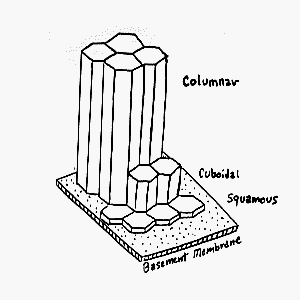
The term squamous is used to describe a cell that is wider than it is
high. Squamous epithelium can be of two types: simple and stratified.
Simple squamous epithelium consists of a single layer of cells.
Here is a simple model that shows how the cells of squamous epithelium
fit together; note the typical hexagonal shape
http://www.udel.edu/Biology/Wags/histopage/modelspage/simpepi.GIF and
here is a comparison of the heights of cells of in different types of
epithelia http://www.udel.edu/Biology/Wags/histopage/EPITHELIAL_HEIGHTS.gif
(both from University of Delaware, resources for course in mammalian
histology)
The nucleus of a squamous epithelial cell will be more like a flattish
disc than like a sphere. Here is another comparative diagram showing
the nuclei: http://webanatomy.net/histology/epithelium/img003.jpg
Examples of simple squamous epithelium:
Walls of blood capillaries
http://webanatomy.net/histology/epithelium/simple_squamous_capillary.jpg
Mesothelium of the peritoneal cavity
http://webanatomy.net/histology/epithelium/mesothelium.jpg
Wall of kidney capsule
http://webanatomy.net/histology/epithelium/ex_simple_squamous.jpg
(on this picture you can see the single row of epithelial cells, with
the nuclei forming “bumps” that rise out of the flat cells)
All these diagrams are from: These diagrams are from “Online anatomy
and physiology resources” by Professor Jim Swan, University of New
Mexico” The whole section on epithelial histology on Jim Swan’s web
site can be accessed from
http://webanatomy.net/histology/epithelial_histology.htm It has a
wealth of information on other types of epithelial tissue as well.
Epithelial cells are joined together by junctions, of which there are
four types:
Tight junctions or “zona occludens” are where the membranes of the
adjacent cells become fused together. The membranes may have many
fusion sites within the junction and so form a seal which prevents the
passage of material through the epithelium. This is what gives the
epithelium its barrier function to prevent things entering the body;
the skin is a good example of this. Sometimes, however, the junctions
are restricted to one part of the cell boundary and so form an
incomplete seal which is called a “fascia occludens”, eg in the
epithelium of blood vessels.
Desmosomes (also called macula adherens) show up as disc shapes on the
surface of a cell, which are matched by the same structure on the
adjacent cell. These discs are attachment sites for bundles of
tonofilaments (made of keratin). These junctions enable the cells to
be held together very firmly.
(NB This is the only type of junction seen in the stratified squamous
epithelium of the skin.)
This is an adhering junction.
Another type of adhering junction is called the zonula adherens. It
looks like a belt around the perimeter of the cell. It is found just
below the tight junctions. The gap between two adjoining cells is
filled with small filaments.
“Together, the zonula occludens, zonula adherens, and macula adherens
constitute the junctional complex visible in the electron microscope.
In the light microscope, the junctional complex is seen as the
terminal bar .”
Gap junctions (also called nexuses) allow communication between cells,
by enabling the passage of cell messenger molecules, some hormones,
ions etc from one cell to another. The membranes of the adjacent
cells come very close, and the tiny gap between them is bridged by
tiny tubular channels which form the pores between the cells through
which substances can pass. These tubes are called connexons. They
are made of protein hexamers with a hydrophillic central pore.
Information on junctions from:
http://www.e-histology.net/index.html#junctions (Altrius Biomedical
Network, E-histology.net) and
http://anatomy.iupui.edu/courses/histo_D502/D502f02/Epithelia/Epithelia.html
(Dept. Anatomy and Cell Biology, University of Indiana Medical School)
Here is a diagram of typical epithelial cell structure, showing the
junctions http://webanatomy.net/histology/epithelial_histology.htm
and a close-up diagram of three of these junctions here
http://webanatomy.net/anatomy/cell_junctions.jpg Please note that
the pictures do not represent a squamous cell as such, but just a
“typical” epithelial cell. A squamous cell is much flatter. However,
otherwise it has the same characteristics as this example.
(From Professor Jim Swan, University of New Mexico, see above for full
details of this site)
I hope this answer is satisfactory, but please ask for further
clarification if required. |
