Hello Spamit,
?Programmed cell death is characterized by the activation of a set
of intracellular cysteine proteases, termed caspases, that cleave
target proteins to cause characteristic morphologic changes associated
with cell death. These changes include nuclear condensation, which is
a prominent feature of erythroblast maturation prior to their
enucleation (see figure). In fact, red cells are not the only
enucleated terminally differentiated cells in mammals.?
?Is there a role for programmed cell death in erythropoiesis?
Immature erythroid cells, particularly at the erythroid colony-forming
unit (CFU-E) and proerythroblast stages, are highly dependent on the
hormone erythropoietin for survival and undergo apoptosis upon
cytokine withdrawal (see figure). This is thought to be an important
mechanism for the regulation of steady-state levels of red cells and
in the "stress" response following acute blood loss, and it likely
involves caspase cleavage of target proteins, including GATA-1.2
However, caspases may also have nonapoptotic roles in erythropoiesis,
since knock-down of caspase-3 transcript levels in erythroid
burst-forming unit (BFU-E) leads to a block in erythroid maturation.3
Little is known about the potential nonapoptotic functions of the cell
death pathway. Caspases are also expressed in aged erythrocytes, but
they do not appear to play a role in the "death" of senescent red
cells.4 Thus, paradoxically, caspases appear to function at an early
stage of erythroid differentiation but not when erythroblasts undergo
the changes in nuclear morphology associated with programmed cell
death. It is becoming ever more apparent that the processes of nuclear
condensation and enucleation that lead to the mature mammalian
erythrocyte are unique in the animal kingdom.?
http://www.bloodjournal.org/cgi/content/full/106/6/1900
?Caspases are a family of cysteine proteases that cleave proteins
after aspartic acid residues. They are the main effectors of apoptosis
or programmed cell death (PCD) and their activation leads to
characteristic morphological changes of the cell such as shrinkage,
chromatin condensation, DNA fragmentation and plasma membrane
blebbing. Induction to commit suicide is required for proper
organismal development, to remove cells that pose a threat to the
organism (e.g. cell infected with virus, cancer cells), and to remove
cells that have damaged DNA. Cells undergoing apoptosis are eventually
removed by phagocytosis.?
http://www.caspases.com/
?Caspases are the executioners of apoptosis. They are protein-cutting
enzymes that chop up strategic proteins in the cell. The name refers
to two properties of these enzymes. First, they are cysteine proteases
that use the sulfur atom in cysteine to perform the cleavage reaction.
Second, they cut proteins next to aspartate amino acids in their
chains. They do not cut indiscriminately--instead, they are designed
to make exactly the right cuts needed to disassemble the cell in an
orderly manner.?
http://www.rcsb.org/pdb/molecules/pdb56_1.html
?Caspases are designed to break proteins into bite-sized pieces, but
the cell needs help to break down its other molecules. Cells also have
a number of caspase-activated proteins to do this work.?
http://www.rcsb.org/pdb/molecules/pdb56_2.html
You may have access to these journals at the MIT library:
----------------------------------------------------------
Involvement of Proteases in Cytokine-Induced Hematopoietic Stem Cell Mobilization
http://www.annalsnyas.org/cgi/content/full/1044/1/60
Apoptotic Role of Fas/Fas Ligand System in the Regulation of Erythropoiesis
http://www.bloodjournal.org/cgi/content/full/93/3/796
Here is a cached page of an article on PCD
Programmed Cell Death
http://scholar.google.com/scholar?hl=en&lr=&c2coff=1&q=cache:5JMZySoqWtoJ:phm.bio.msu.ru/edocs/samuilov-2-en.pdf+erythrocyte++%2B+marrow+%2B+cysteine+proteases+%2B+nucleus
RBC Maturation
===============
?Reticulocytes, on their way to becoming mature RBCs, leave the bone
marrow. They contain remnant RNA which stains blue on the pink
cytoplasm (polychromasia). They squeeze between the endothelial cells
to enter the sinusoid, leaving their nuclei behind. The debris from
the maturing RBC including the iron is then picked up and recycled by
a nurse cell.?
As a red cell matures, the nucleus undergoes karyorrhexis, meaning the
nucleus degrades. The nuclear chromatin is dispersed into the
cytoplasm of the cell. These fragments then escape the cell.
http://medical-dictionary.thefreedictionary.com/karyorrhexis
This site has a great illustration:
?The cell is shown extruding its nucleus to become an immature
erythrocyte (a reticulocyte), which then leaves the bone marrow and
passes into the bloodstream. The reticulocyte will lose its
mitochondria and ribosomes within a day or two to become a mature
erythrocyte. Erythrocyte clones develop in the bone marrow on the
surface of a macrophage, which phagocytoses and digests the nuclei
discarded by the erythroblasts.?
http://www.ncbi.nlm.nih.gov/books/bv.fcgi?rid=mboc4.figgrp.4158
?Finally, the cell extrudes its nucleus, but some polyribosomes are
retained in the cell which still produce hemoglobin. Cells at this
stage are identifed using special stains that cause the ribosomes to
clump together. These cells are called reticulocytes, and normally
they make up 1-2% of the red blood cells found in the blood stream.?
http://cats.med.uvm.edu/cats_teachingmod/histology/lectures_online/blood_and_hemat/erythropoiesis/history.html
A mature, circulating red blood cell (erythrocyte) has no nucleus (and
no DNA) ? the nuclear chromatin (RNA) has been absorbed by the RBC.
?Colored or red corpuscles (erythrocytes), when examined under the
microscope, are seen to be circular disks, biconcave in profile. The
disk has no nucleus, but, in consequence of its biconcave shape,
presents, according to the alterations of focus under an ordinary high
power, a central part, sometimes bright, sometimes dark, which has the
appearance of a nucleus (Fig. 453, a). It is to the aggregation of the
red corpuscles that the blood owes its red hue, although when examined
by transmitted light their color appears to be only a faint reddish
yellow.?
http://www.bartleby.com/107/134.html
?Above age 20, most RBCs are produced primarily in the marrow of the
vertebrae, the sternum, the ribs, and the pelvis. Let's examine how
RBCs are produced and, ultimately, how they are destroyed.?
?In the bone marrow there are many special stem cells from which RBCs
can be formed. As these cells mature, they extrude their nucleus as
they slowly fill with hemoglobin until they are bright red
reticulocytes ready to escape the bone marrow and squeeze into the
blood capillaries to begin circulating around the body. In a blood
sample, the reticulocytes can be distinguished from RBCs because they
still contain some speckles or pieces of their nucleus. Within a few
days, this reticulocyte completely loses all its nuclear material and
becomes a full-fledged RBC that is ready to serve the oxygen needs of
the body. After about three to four months, the RBC has worked so hard
that it begins to weaken. The membranes of old RBCs become very
fragile and the cells may rupture during passage through some tight
spots in the circulation. These old and damaged RBCs are "eaten"
primarily by the spleen, and most of the leftover components
(especially the iron from the hemoglobin) are recycled to form new
RBCs.?
http://www.nsbri.org/HumanPhysSpace/focus3/erythropoiesis.html
The process of RBC maturation: A red cell starts as a stem cell, and
becomes an erythrocyte (RBC) in about 120 hours.
http://campus.murraystate.edu/academic/faculty/Wade.Northington/Lecture%20Notes%20PDF%20Format/Erythrocyte%20Maturation.PDF
?Red cells become smaller as they mature. Their cytoplasm starts off
as deep blue due to a high RNA content and becomes purplish blue as
hemoglobin production begins. When the cell is mature and fully
hemoglobinated, the cytoplasm stains pink with Wright's stain. The
young nucleus is dark red in color and has fine lacy chromatin.
Nucleoli may be seen. As the cell matures the nucleus condenses,
stains dark bluish-purple and is eventually extruded from the cell.
Nucleoli are present in the young red cell and absent in the mature
red cell. The light spot in the cytoplasm (adjacent to the nucleus)
represents the golgi apparatus and is prominent in young cells,
becoming non-distinct as the cell matures.?
http://www.academic.marist.edu/~jzmz/HematologyI/NRBC7.html
?Erythropoeitin stimulates red cell precursors at all levels of
maturation to hasten the maturation process. It is also responsible
for stimulating the premature release of reticulocytes into the
bloodstream. As RBCs mature, they gradually lose their membrane
erythropoeitin receptors (and therefore their ability to respond to
erythropoeitin) until they are left with none at the mature
erythrocyte stage.?
?Reticulocytes retain RNA and ribosomes. With RNA the reticulocyte
continues to produce hemoglobin. As the reticulocyte transforms into a
mature RBC it gradually loses its RNA; as a result the hemoglobin
synthesizing potential gradually decreases until the cell no longer
produces any hemoglobin. The mature red cell has all the hemoglobin it
will carry for its entire 120-day lifespan. The reticulocyte can
produce up to 30% of the body's total hemoglobin stores. (The other
70-80% is made in the pre-reticulocyte stages).?
http://edcenter.med.cornell.edu/CUMC_PathNotes/Hematopathology/Hematopathology.html
�?Cell volume decreases as the cell matures. On the average the size
goes from rubriblast measuring 12-19 microns to a mature erythrocyte
measuring 6-8 microns in diameter.
�Chromatin condenses. The rubriblast has very fine chromatin; the
metarubricyte has solid chromatin and the mature erythrocyte has none.
�Nucleoli disappear by the rubricyte state.
�Nuclear shape remains round.
�N:C ratio decreases. The rubriblast and prorubricyte have an N:C
ratio of 4:1; the rubricyte and metarubricyte N:C ratio is 1:1.
�RNA activity decreases in cytoplasm resulting in lighter blue
cytoplasm as the cell matures. The rubriblast cytoplasm is deep blue,
the rubricyte cytoplasm is pinkish blue, the pink coming from the
beginning of hemoglobin production.
�Hemoglobin production begins at the rubricyte stage and increases as
the cell matures. There is a gradual shifting of predominantly blue to
predominantly pink cytoplasmic color as the cell matures to an
erythrocyte. A mature erythrocyte has no blue color in the cytoplasm.
�Mitochondria activity decreases. You may see a halo around the
nucleus in the rubriblast and prorubricyte.
�The nucleus is eventually extruded. The metarubricyte is last stage
with a nucleus.
�Blue color of cytoplasm due to presence of RNA indicating protein synthesis.
�Pink color of cytoplasm due to presence of hemoglobin production.
�Perinuclear halo indicates mitochondria and Golgi apparatus surround
the nucleus. These structures do not pick up stain.
�From 14 to16 erythrocytes are produced from one rubriblast.
http://www.ndsu.nodak.edu/instruct/tcolvill/435/erythrocytes.htm
In certain diseases, one can see reticulocytes in the circulating blood.
?Reticulocytes are immature red blood cells (RBC) which have shed
their nucleus, but still retain residual nuclear material. Clinically,
the reticulocyte percentage is a useful indicator of erythropoiesis.
In cases of anaemia, an elevated reticulocyte count is indicative of
normal marrow function, whilst a decreased result may be more
consistent with impaired erythropoiesis. Traditionally the
reticulocyte percentage was estimated by precipitating the residual
RNA with a dye, and counting the stained cells as a percentage of 1000
RBC using a microscope. This method is well known to be imprecise and
open to subjective interpretation by the technologist.?
http://jcsmr.anu.edu.au/facslab/AFCG/Standards/reticulo.html
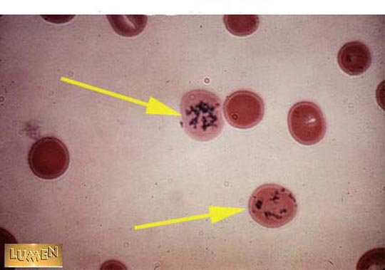
?Nuclear fragments. In some disease states, nuclear fragments, or
Howell-Joliy bodies, remain in otherwise mature RBCs. When these form
circular tilaments they are termed Cabot rings. c. Reticulocytes. Some
RBCs recently released from the bone marrow contain a small amount of
residual RER and ribosomes that can be precipitated into blue, netlike
struc tures with the vital dye brilliant cresyl blue. When these
reticulocytes constitute more than about 1% of the circulating RBCs,
they indicate an increased demand for oxygen carrying capacity leg,
from loss of RBCs due to hemorrhage or anemia, or to recent ascent to
a higher altitude)?
http://www.loyno.edu/~chood/histnotesbloodhem.html
?The orthochromatic normoblast extrudes its nucleus leaving behind a
reticulocyte. The extruded but functionally impaired nucleus of the
orthochromatic normoblast then gives rise to a polychromatic
normoblast, a defective cell. The poorly made cytoplasm of the
polychromatic normoblast is shed and its nucleus, now non-functional,
undergoes complete dissolution into an aggregate of ultrafine
particles.?
http://www.ncbi.nlm.nih.gov/entrez/query.fcgi?cmd=Retrieve&db=PubMed&list_uids=7666825&dopt=Abstract
Ilustrations:
=============
Nucleus being extruded
http://www.academic.marist.edu/~jzmz/HematologyI/NRBC13.html
Click the small red box to see a slide show of bone marrow sinuses
http://ect.downstate.edu/courseware/histomanual/hematopoiesis.html
Scroll down to B: Erythropoeisis and click on Illustration 1, to see
the whol maturation process of an RBC. Scroll down to C: and click
onto Illustration. This is a white blood cell phagocytosing an old
RBC.
http://campus.murraystate.edu/academic/faculty/Wade.Northington/erythroc1.htm
Scroll down to slide 99600 and b.12
http://medic.med.uth.tmc.edu/edprog/histolog/blood/hist-08.htm
An image of reticulocytes
http://www.irvingcrowley.com/cls/retic.jpg
Another
http://www.lumen.luc.edu/lumen/meded/mech/cases/case7/hp5-15.jpg
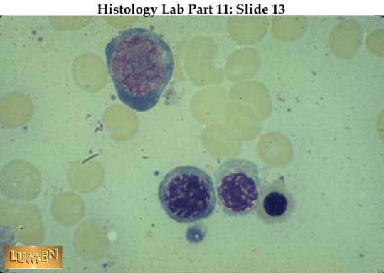
Immature RBCs. The bottom row includes a basophilic normoblast
(erythroblast), and a polychromatophilic normoblast (erythroblast) and
an orthochromatic (or eosinophilic) normoblast.
http://www.lumen.luc.edu/lumen/meded/mech/cases/case7/hl5-13.jpg
I hope this has helped you out! If any part of this answer is unclear,
please do not rate it. Simply ask for an Answer Clarification, and I
will be happy to assist you further, before you rate this answer.
Regards, Crabcakes
Search Terms
=============
erythropoeisis
Human reticulocytes
Human erythrocyte nucleus
Human erythrocyte maturation
red cell nucleus extruded + phagocytosis |
Clarification of Answer by
crabcakes-ga
on
01 Oct 2005 11:07 PDT
Hello again Spamit,

   Thank you for your clarification. I was unable to discern from your
question exactly which level of information you were seeking.

I believe the following should be more informative:

?The main regulator of that process is erythropoietin, a glycoprotein
hormone that circulates at about one hundredth of the concentration of
most other hormones in the body [1, 2]. Erythropoietin is produced in
the kidneys. It circulates in the plasma and induces red cell
production in the bone marrow [3], where it binds to erythroid
progenitor cells. Cell culture studies have identified two classes of
erythroid progenitor cells, BFU-E and colony forming units-erythroid
(CFU-E) (Fig. 1 ). Both types of cell have receptors for
erythropoietin on their surfaces. When erythropoietin binds to its
receptors on BFU-E cells, they proliferate into CFU-E
(proerythroblasts). Proerythroblasts are exquisitely sensitive to
erythropoietin. They proliferate and develop into erythroblasts and
then reticulocytes that enter the peripheral circulation where they
mature into red blood cells.

Erythropoietin is a glycoprotein molecule composed of 165 amino acids
and four carbohydrate groups. The primary structure is shown in Figure
2 . An important structural feature of erythropoietin is that it has
two disulphide bonds, one linking the cysteine at amino acid 6 with
the cysteine at amino acid 161, and the other linking cysteines 29 and
33. The former is functionally more important, because it acts as a
tether, ensuring that the whole molecule is held in the correct shape
for binding to the erythropoietin receptor. If this bond breaks, the
molecule loses its biological activity. From the structure of the
erythropoietin molecule, and from experimental evidence, one can infer
that molecules of erythropoietin aggregate together through a process
known as hydrophobic interaction. When this happens to any degree,
perhaps as a result of improper storage, erythropoietin becomes
significantly less potent.?
http://theoncologist.alphamedpress.org/cgi/content/full/8/suppl_1/15

This is a cached page (It may not be online much longer)
?The main stages of erythrocyte maturation are regulated by the gene
network described in GeneNet database. The hormone erythropoietin
interacts with immature erythroid cells (erythroid stem progenitors of
CFU-E type) and stimulates their proliferation, syntheses of
hemoglobin, and the enzymes involved in heme biosynthesis, that is,
maturation and differentiation of erythroid progenitors. Low partial
pressure of oxygen in venous blood (hypoxia) is another stimulator of
erythropoietin synthesis.?
http://66.102.7.104/search?q=cache:43fx31VdXmUJ:wwwmgs.bionet.nsc.ru/mgs/gnw/gn_model/5.shtml+Erythropoietin++%2B+erythrocyte+maturation&hl=en


?p38 MAP kinase (p38) and JNK have been described as playing a
critical role in the response to a variety of environmental stresses
and proinflammatory cytokines. It was recently reported that
hematopoietic cytokines activate not only classical MAP kinases (ERK),
but also p38 and JNK. However, the physiological function of these
kinases in hematopoiesis remains obscure. We found that all MAP
kinases examined, ERK1, ERK2, p38, JNK1, and JNK2, were rapidly and
transiently activated by erythropoietin (Epo) stimulation in SKT6
cells, which can be induced to differentiate into hemoglobinized cells
in response to Epo. Furthermore, p38-specific inhibitor SB203580 but
not MEK-specific inhibitor PD98059 significantly suppressed
Epo-induced differentiation and antisense oligonucleotides of p38,
JNK1, and JNK2, but neither ERK1 nor ERK2 clearly inhibited
Epo-induced hemoglobinization. However, in Epo-dependent FD-EPO cells,
inhibition of either ERKs, p38, or JNKs suppressed cell growth.
Furthermore, forced expression of a gain-of-function MKK6 mutant,
which specifically activated p38, induced hemoglobinization of SKT6
cells without Epo. These results indicate that activation of p38 and
JNKs but not of ERKs is required for Epo-induced erythroid
differentiation of SKT6 cells, whereas all of these kinases are
involved in Epo-induced mitogenesis of FD-EPO cells.?
http://www.bloodjournal.org/cgi/content/full/92/6/1859

http://stemcells.alphamedpress.org/cgi/content/full/18/5/366


Erythropoietin is a glycoprotein. It acts on the bone marrow to
increase the production of red blood cells. Stimuli such as bleeding
or moving to high altitudes (where oxygen is scarcer) trigger the
release of EPO.
http://users.rcn.com/jkimball.ma.ultranet/BiologyPages/K/KidneyHormones.html#epo


Growth hormone and erythropoietin differentially activate DNA-binding
proteins by tyrosine phosphorylation.
http://www.pubmedcentral.nih.gov/articlerender.fcgi?artid=358571&tools=bot


You can register at this site to be able to purchase this article:
?Erythropoietin has been the fountainhead in research on molecular
mechanisms of oxygen-sensing and hypoxia-induced gene expression. The
availability of recombinant human EPO for prevention and therapy of
chronic anemias was a turning point in clinical medicine. This book
intends to provide a fairly complete overview and bibliography of the
progress made in important areas of basic and clinical EPO research.
It is divided into sections on EPO structure and control of
production; EPO mechanisms of action; EPO physiology and
pathophysiology; EPO pharmacology and therapy; and recombinant human
EPO therapy.?
http://www.sigmaaldrich.com/catalog/search/ProductDetail?ProdNo=Z700703&Brand=SIGMA


?Developing erythroid precursor cells in the bone marrow express
erythropoietin receptors at about the BFU-E stage of maturation. The
cells each express, at most, 3-400 erythropoietin receptors. In
response to erythropoietin and a number of other erythropoietic
stimulatory hormones, the red cell precursors produce mature
erythrocytes (Figure 2) (Adamson, 1994).?
http://sickle.bwh.harvard.edu/iron_epo.html

More References
http://www.fpgrahamco.com/Pages/054.html

I quoted an article about apoptosis because apoptosis is a component
of normal erythropoiesis, and is being researched.
?In the absence of erythropoietin, erythroid progenitor cells
accumulated DNA cleavage fragments characteristic of those found in
programmed cell death (apoptosis) by 2 to 4 hours and began dying by
16 hours. In the presence of erythropoietin, the progenitor cells
survived and differentiated into reticulocytes. Thus, apoptosis is a
major component of normal erythropoiesis, and erythropoietin controls
erythrocyte production by retarding DNA breakdown and preventing
apoptosis in erythroid progenitor cells.?
http://www.ncbi.nlm.nih.gov/entrez/query.fcgi?cmd=Retrieve&db=PubMed&list_uids=2326648&dopt=Citation

Hope this additional information has further helped you.

Regards, Crabcakes
|
